Egyptian mandarin peel oil's anti-scabies potential via downregulation-of-inflammatory/immune-cross-talk: GC–MS and ... - Nature.com
Abstract
The current study investigated the scabicidal potential of Egyptian mandarin peel oil (Citrus reticulata Blanco, F. Rutaceae) against sarcoptic mange-in-rabbits. Analysis of the oil's GC–MS identified a total of 20 compounds, accounting for 98.91% of all compounds found. Mandarin peel oil topical application improved all signs of infection, causing a scabicidal effect three days later, whereas in vitro application caused complete mite mortality one day later. In comparison to ivermectin, histopathological analysis showed that the epidermis' inflammatory-infiltration/hyperkeratosis-had disappeared. In addition to TIMP-1, the results of the mRNA gene expression analysis showed upregulation of I-CAM-1-and-KGF and downregulation of ILs-1, 6, 10, VEGF, MMP-9, and MCP-1. The scabies network was constructed and subjected to a comprehensive bioinformatic evaluation. TNF-, IL-1B, and IL-6, the top three hub protein-coding genes, have been identified as key therapeutic targets for scabies. From molecular docking data, compounds 15 and 16 acquired sufficient affinity towards the three screened proteins, particularly both possessing higher affinity towards the IL-6 receptor. Interestingly, it achieved a higher binding energy score than the ligand of the docked protein rather than displaying proper binding interactions like those of the ligand. Meanwhile, geraniol (15) showed the highest affinity towards the GST protein, suggesting its contribution to the acaricidal effect of the extract. The subsequent, MD simulations revealed that geraniol can achieve stable binding inside the binding site of both GST and IL-6. Our findings collectively revealed the scabicidal ability of mandarin peel extract for the first time, paving the way for an efficient, economical, and environmentally friendly herbal alternative for treating rabbits with Sarcoptes mange.
Microbiota in health and diseases
Small molecule induced STING degradation facilitated by the HECT ligase HERC4

Complete biosynthesis of QS-21 in engineered yeast
Introduction
Sarcoptic mange (Sarcoptes scabiei) is a serious infectious disease that invades humans and animals all over the world1. The mites are highly adapted to contact with their host as contagious, burrowing, and obligate parasites. Sarcoptic mange Grower pig production is negatively impacted by adult female mites; because they mate on the skin's surface, burrow into the skin, lay eggs, and cause irritations that can lead to bleeding, reduced feeding and development, chronic stress, and decreased welfare2,3. The clinical picture represents chronic hyperkeratotic, which is characterized by the presence of aural crusts and many mites on the animal4. Similar to people, rabbits are susceptible to Sarcoptes infection, or mange, which reduces production and causes economic losses for rabbits, especially in the absence of effective treatment5. Therapy options include the systemic treatment of macrocyclic lactones, local administration of amitraz or pyrethroids, or both6,7. Despite their long history of effectiveness in treating mange, their extensive use has led to a decline in effectiveness because of the emergence of drug resistance. Thus, it is crucial to create novel scabicides that are both efficient and secure in order to treat and control mammalian scabies6.
In rabbits, goats, and pigs, several essential oils derived from Citrus limon, Lavandula angustifolia, Citrus aurantium amara, Pelargonium asperum, Melaleuca alternifolia, Syzygium aromaticum, Eucalyptus radiata, Leptospermum scoparium, Juniperus oxycedrus, Cryptomeria japonica, and Cymbopogon martini, were put to the test in real time against S. scabiei8,9,10,11,12. Essential oils are typically favoured over chemical acaricides since they are less harmful to animals and have a shorter environmental persistence. Also, the complex chemistry of essential oils is known to considerably impede the emergence of drug resistance against these chemicals13. Yet, because essential oils consist of a complex mixture of components, it might be challenging to attribute an essential oil's acaricidal properties to a specific ingredient or combination of compounds14. Skin irritation is yet another potential drawback that has been reported in humans15.
Some of the most coveted Citrus fruits for fresh consumption are mandarins, C. reticulata16. The more frequent name for them is "mandarin," but they are also occasionally called "tangerines." The Mandarin species includes a number of cultivars and hybrids16. Popularly grown varieties include C. unshiu Marcovitch (also known as Unshiu mikan in Japanese), C. nobilis Loureiro (also known as king mandarins), C. deliciosa Tenore (also known as Mediterranean mandarins), and C. reticulata Blanco (common mandarins)16,17. Mandarins are one of the main Citrus fruits grown in many countries such as China, Brazil, USA, India, Mexico, Spain, etc. The fruits have a great commercial worth for their essential oils and other fragrant compounds, even though they are primarily used to make pastries18. A lot of beverages, candies, cookies, and desserts use Citrus flavours19, while the peels of C. reticulata are used to flavour alcohol19. Citrus reticulata EO shown an anti-proliferative activity against rat pulmonary fibrosis produced by bleomycin (BLM) and protective properties against human embryonic lung fibroblasts (HELFs). The method is believed to involve correcting the imbalance between oxidation and antioxidation, lowering collagen deposition and fibrosis, and down-regulating lung tissue expressions of connective tissue growth factor (CTGF) and mRNA20. Due to its high d-limonene concentration21, C. reticulata EO demonstrated a moderate level of radical scavenging action22. Mandarin oil is well known for its broad spectrum antibacterial and antifungal actions. It inhibits the growth of several bacteria including Escherichia coli, Bacillus subtilis, Pseudomonas aeruginosa, and Staphylococcus aureus22,23, as well as several fungi including Penicillium italicum, P. chrysogenum, P. digitatum, Aspergillus niger,-A. flavus, Alternaria alternata, Curvularia lunata, Rhizoctonia solani, Fusarium oxysporum, and-Helminthosporium oryzae23,24,25,26.
The GC–MS profiling of mandarin peel oil has been used in the current study. Additionally, for the first time, through-in vitro,-in vivo,-histopathology,-mRNA-expression, and network/in silico analysis, the extract's scabicidal potential against-Sarcoptic-mange-in-rabbits has been investigated, allowing for the incorporation of natural candidates to proper and secure management of infectious diseases. The present investigation's framework is shown in Fig. 1.

General outflow of the study.
Material and methods
Ethical permission
Plant materials and experiments were conducted in accordance with relevant institutional, national, and international guidelines. The study took place according to the ethical committee's permission number of 9/5/2022 at Deraya College. It was done in accordance with the National Institute of Health's guidelines for the care and use of laboratory animals and ARRIVE guidelines27.
Fruit collection
In January 2021, C. reticulata cultivated fruits were harvested from a house garden on Atia Street in Beni-Suef, Egypt. A voucher specimen (2021-BuPD-88) was deposited at Pharmacognosy-Department, Faculty-of-Pharmacy, Beni-Suef-University, Egypt.
Sample preparation
Using the Clevenger apparatus, the fresh peels (0.5 kg) were hydrodistillated for two hours at 75 °C. The oil was gathered, dried over anhydrous sodium sulphate, and kept in airtight amber glass vials at 4 °C for storage. On the basis of the plant material's fresh weight, the yield (v/w%) was computed28,29.
GC–MS analysis
Gas chromatography-mass spectrometry (GC/MS) was used to perform chromatographic analysis on the oil recovered from peels28,30. The GC–MS apparatus combines a thermal mass spectrometer detector (ISQ single quadrupole mass spectrometry) with a TRACE GC ultra-high performance gas chromatograph (THERMO Scientific Corp., USA). A TR-5 MS column (30 m × 0.32 mm i.d., 0.25 mm film thickness) was installed in the GC–MS system. For the analyses, He-lium was used as the carrier gas, and the split ratio was set at 1:10 using the following temperature program: 60 C for 1 min, followed by 4.0 C/min to 240 C and a 1-min hold. At 210 °C, the injector and detector were maintained. One-liter samples of the mixes were always administered as diluted samples (1:10 hexane, v/v). By using a spectral range of m/z 40–450 and electron ionization (EI) at 70 eV, mass spectra were produced. Using AMDIS software (www.amdis.net), the chemical components of the essential oil were deconvoluted and identified by their retention indices (relative to n-alkanes C8-C22), mass spectra matching to genuine standards, and retention times (when available). (NIST Standard Reference Database, 78 Version 5.10) Wiley spectral library collection28,31,32.
In vitro assays
In-vitro-antioxidant-activity
Hydrogen-peroxide-scavenging-activity
The reaction with a defined amount of exogenously provided hydrogen peroxide (H2O2) was used to determine H2O2 scavenging activity that reflects the anti-oxidative capacity of the peel oil. Colorimetric analysis was used to estimate the residual H2O233. In brief, 20 µl of the sample was mixed with 500 µl of H2O2 and incubated at 37 °C for 10 min. 500 l of the enzyme/3, 5-dichloro-2-hydroxyl-benzensulfonate solution were then added, and it was incubated at 37 °C for 5 min. The colored product's intensity was quantified colorimetrically at-510-nm. A positive control was ascorbic acid. By comparing the test results to those of the control group, the percentage-of H2O2-scavenging activity-was calculated and applying the following formula:
IC50 of each sample was calculated after performing the assay at eight different concentrations : (1000, 750, 500, 375, 250, 187.5, 125 and 0 µg/mL) using Graph pad prism 7 software.
Superoxide radical scavenging activity
The scavenging activity of superoxide anion was measured34. In a Tris-HCL solution (16 mM, pH 8.0) containing 90 l of NBT (0.3 mM), 90 l of NADH (0.936 mM), 0.1 ml of peel oil (125, 250, 500, and 1000 g/mL), and 0.8 ml of Tris-HCl buffer, superoxide anion radicals were produced (16 mM, pH 8.0). After adding 0.1 ml of PMS solution (0.12 mM) to the mixture, the reaction was started. The mixture was then incubated at 25 °C for 5 min, during which time the absorbance was measured at 560 nm. Ascorbic acid was used as a model substance. Using the formula below, the percentage inhibition was calculated by comparing the test results to those of the control:
IC50 was estimated by doing the test at four different concentrations and using the GraphPad Prism 7 software.
Biological investigation
Collection-of-Sarcoptes-scabiei-mites
Adult-mites-were collected from rabbits that were-naturally-infected, Deraya University, Minia, Egypt's Animal House. Scraped from the borders of the lesions, the infected skin samples were then shifted-to-petri plates and-incubated within a biochemical-oxygen-demand (BOD) for an incubator for 30 min at 35 °C.
In vitro-application of peels oil on-sarcoptic-mange
A petri dish containing mites was filled with 2 ml of diluting extract (20%), along with the plates were then incubated-in-BOD. Reaction observations were made-at 1, 12, and 24 h after application. Petri plates were incubated at an ambient temperature of 25 °C and with a relative moisture of 75%, with a 5% ivermectin (1 cm3/l) group as the positive-control-and distilled water as the-negative-control. By stimulating the mites with a needle, the death of the mite was confirmed; the mite was deemed dead if it showed no response.
In vivo application of peels oil
The study took place on male adult rabbits for 4 weeks (weighing 2.8–3.2 kg) that were infected. The animals' ears showed clinical indicators of mange infection, such as hyperkeratinization, inflammation, redness, itching, and irritability. Microscopic mite identification in skin scrapings further corroborated this. Four groups of five rabbits each were made up of twenty animals, as follows: Five rabbits made up the normal group, the paraffin oil-positive-control-group. The-ivermectin-treated-group (5%-ivermectin). The peel-oil-group (20%-peel-oil in paraffin-oil). Paraffin oil, which is a-mineral-oil, was reportedly chosen as a diluent for the peel oil because it has little impact-on-mites35. Each group were kept in a separate cage, and each group received treatment by dipping the infected ears once daily. Steel hoppers were used to feed all of the rabbits, and water was available at all times. The rabbits were observed every two days to assess their clinical recovery. The goal was to find any signs of improvement in the lesions, such as the absence of irritation and redness, cutaneous smoothing, the start of the development of hair from the infection, and the cessation of scab development10. Skin scrapings from each rabbit's sick and healed areas were taken every three days, and Throughout the course of the therapy, they were microscopically investigated to check for sarcoptic mites with a LEICA, DM1000 microscope with a digital camera (LEICA, EC3, Germany)10.
Histopathological-examination
Tissue samples were collected at zero and three weeks following the start of the course of therapy via 20% peeling oil as well as ivermectin from healthy and infected ears. Following that, samples were dried in ethyl alcohols of increasing strength, sterilized via xylene, infused with paraffin that had been melted at 55–60 °C, and finally inserted into paraffin wax. The samples were then preserved in 10% buffered formalin. Deparaffinized, rehydrated, and stained with hematoxylin and eosin (H & E), "3–5 m thick" tissue sections were examined using-a-light-electron-microscope36.
RNA-isolation-and-qRT-PCR-assay
Using-a-digital-homogenizer-(Branson-Digital-Homogenizer®,-Danbury,-CT,-USA), 100 mg of the tissues under investigation were homogenised in 1 ml of TRIzolTM RNA Extraction Reagent (Amresco, Solon, OH, USA). RNA extraction from the biopsy sample was done in accordance with the manufacturer's instructions. RevertAid H-minus First Strand cDNA Synthesis Kits (#K1632, Thermo Science Fermentas, St. Leon-Ro, Germany) were used to create cDNA from the extracted RNA for comparable amounts of total RNA in all samples. The-qRT-PCR-was carried out on-the-Applied-Biosystems Step One Plus system using the cDNA as a template. The primers were created using the NCBI primer blast software and were produced by Invitrogen. Using the GAPDH gene as a housekeeping gene, data were analyzed using the 2CT approach31. Table 1 lists the primer sequences that were employed.
In silico studies
Construction of protein–protein interaction (PPI) network
Using Cytoscape 3.9.1 software (https://www.cytoscape.org/)37 and by lunching STRING disease query tool incorporated in it which retrieves network for the top human proteins associated with the queried disease from a weekly updated web source of diseases database (https://string-db.org/)38 Scabies was chosen as the search term, and "Human sapiens" was chosen as the species type. The confidence score was set to 0.4, and the default settings for the remaining parameters were used to create the PPI network39.
Hub gene expression analysis
The plugin for cytoHubba the hub genes are identified using ranking techniques such as degree, edge percolated component (EPC), maximum neighbourhood component (MNC), the density of maximum neighborhood component (DMNC), and maximal clique centrality (MCC), as well as bottleneck, eccentricity, closeness, radiality, betweenness, stress, and clustering coefficient. Cytoscape is regarded as a useful exploration interface for the most significant nodes in PPI networks40,41.
Gene ontology and enrichment analysis
We employed a freely accessible bioinformatics web tool in the current investigation (ShinyGO v0.76.3). Using the many bioinformatics databases accessible, it is possible to perform both gene ontology enrichment analysis and pathway enrichment analysis. ShinyGO was used to perform the gene ontology and enrichment analysis on the 16 genes to determine the cellular elements, molecular functions, and biological processes that were impacted by this set of genes. ShinyGO retrieves comprehensive descriptions of biological signal transduction pathways from numerous databases42.
Molecular docking study
The methodologies of molecular docking intend to predict the best binding orientation of a ligand to a receptor. It proposes several suitable poses of the ligand within the active or docking site of a receptor molecule.in this study, twenty compounds that were identified underwent an in silico study by using screening for three different important-protein-targets that are heavily involved in the scabies infection process, as well as screening for potential targets at the mite itself as an acaricidal effect, in an attempt to get deep inside the mechanistic anti-scabietic effect of orange oil. The chosen targets include IL-1, which is highly effective in stimulating T cells with regulatory functions, and IL-6, which is involved in the formation of Th17 lymphocytes and the release of IL-1743. These cy-tokines have been identified as one of the primary molecules responsible for allergic Th2-type inflammation in the immunological response to scabies, along with TNF-, which is significant in alternative macrophage activation44. GSH, which is linked to the scabies defense system, takes part in a variety of processes crucial to the preservation of cells from oxygen and free radical oxidative damage45, Its distinctive anti-oxidant action makes it a potential target for the oil's acaricidal impact46. In our docking investigation, we validated the ligand and visualized the many docked poses using the computer programme MOE 2019.010. TNF- complexed with its ligand (PDB ID code: 2AZ5) is the last one, and GST is the other protein target of the mite delta class. The first protein target is (IL-1), depicted by the protein's PDB ID code of 6Y8M in co-crystallization with IL-6, as reflected by PDB ID code 1ALU, and its inhibiting ligand SX2 (a-bromo-amido-pyridine-derivative)47 represented by proteins (PDB ID code: 3EIN), the selected targets were acquired via the web from the Database of Proteins (http://www.rcsb.org/pdb).
Molecular dynamic simulation
The MD simulations were carried out using NAMD 3.0.0. software48,49. The Charmm-36 force field is implemented in this piece of software. The protein structure was examined for missing hydrogens, the protonation states of the amino acid residues were set (pH = 7.4), and the co-crystallized water molecules were removed using the QwikMD toolkit of the VMD software. The entire assembly was then packed into a 20 solvent buffer containing 0.15 M Na + and Cl- ions in an orthorhombic box of TIP3P water. After 5 ns of equilibration, the systems were subjected to an energy minimization protocol. Force Field Toolkit (ffTK), a plugin for the VMD software, was used to determine ligand properties and topologies. After the parameters and topology files were prepared, they were imported into VMD so that the protein–ligand complexes could be read accurately, and the simulations could be run.
Statistical analysis
The data were tabulated using the statistical programme-GraphPad-Prism-version-9 (GraphPad,-La-Jolla,-CA,-USA). To evaluate statistical differences between the groups, the ANOVA test was performed, followed by the-Bonferroni-post-hoc-test-for multiple-comparisons. The threshold for statistical significance is a p-value of 0.05 or less.
Results
GC–MS profiling of mandarin peels oil
Egyptian C. reticulata peels gave 2.6% v/w volatile oil fresh weight, being colourless with a characteristic odor, lighter than water, clear, transparent, and not viscous at room temperature as well as at 4 °C. GC–MS analysis was used to identify a total of 20 compounds, accounting for 98.91% of all compounds found (Table 2, Figs. 2, 3). The identified compounds 1–20 belonged to different chemical classes, including monoterpene, phenylpropene, fatty alcohol, and sesquiterpene (Table 2, Fig. 3). where monoterpenes represented 92.16% of the total identified compounds, followed by phenylpropene (3.01%), fatty alcohol (2.36%), and sesquiterpene (1.38%) (Table 2). Fourteen monoterpenes compounds (92.16%) were identified; ranging from cyclic hydrocarbon (D-limonene 4, γ-terpinene 6, 73.32%) which represented the major oil fraction, to oxygenated cyclic hydrocarbon ((-)-isomenthone 10, terpinen-4-ol 11, (-)-carvone 14, 3.32%), and oxygenated acyclic hydrocarbon (linalool 8, citronellol 13, geraniol 15, 8.78%), acyclic hydrocarbon (α-myrcene 3, α-ocimene 5, 2.84%), bicyclic hydrocarbon (α-pinene 1, sabinene 2, 3.45%), to oxygenated bicyclic hydrocarbon (camphor 9, 0.45%) (Table 2, Fig. 3). Also, phenylpropene class (3.01%) contained 2.70 and 0.31% of estragole 12, and anethole 17, respectively. The detected fatty alcohol class contained only 1-octanol 7 and 1-decanol 16, representing 2.36% (Table 2, Fig. 3). On the other hand, three sesquiterpene compounds (1.38%) were identified, varying from a bicyclic hydrocarbon (caryophyllene 19, ( +)-valencene 20, 1.00%), to a tricyclic hydrocarbon (α-copaene 18, 0.38%) (Table 2, Fig. 3).

GC/MS spectrum for Citrus reticulata peels oil.
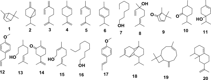
Structures of identified compounds, using GC/MS analysis, from Citrus reticulata oil isolated from peels.
According to the literature, the chemical composition of essential oils varies depending on the age of the plant, harvesting time, geographical location, and environmental conditions50. The Indian C. reticulata peels volatile oil differently from the Egyptian, having 80 compounds, where monoterpene (63.80%), represents mainly limonene (50.42%), myrecene (3.03%), and α-terpineol (1.19%), while sesquiterpene (12.98%) represents mainly α-copaene (1.49%), β-copaene (1.30%), and α-humulene (1.23%). The Indian C. reticulata oil is characterised by its high content of fatty acids (8.73%) and aldehyde content (7.08%), mainly n-hexadecanoic acid (5.65%) α-sinensal (3.14%)51. The essential oil isolated from fully matured, ripened Indian fruit peels of C. reticulata, on the other hand, contained 37 different components (99%). The primary ingredients included limonene (46.7%), geranial (19.0%), neral (14.5%), geranyl acetate (3.9%), geraniol (3.5%), -caryophyllene (2.6%), nerol (2.3%), neryl acetate (1.1%), and others26.
The essential oil constituents reported in C. reticulata grown in Burundi contained 58 constituents52. The most prevalent chemical category was monoterpene hydrocarbons (94.7%). Limonene accounted for 84.8% of the total composition, with -terpinene (5.4%), myrcene (2.2%), and -pinene (1.1%) following. Germacrene D and valencene were the primary components of the sesquiterpene hydrocarbons, which made up only 0.2% of the total composition. Compounds containing oxygen from different chemical groups made up 2.3%52. The two main chemical groupings were terpene alcohols (0.7%) and aliphatic aldehydes (0.7%). Linalool (0.7%), octanal (0.5%), and decanal (0.2%) made up the bulk of the mixture. In concentrations of 0.1%, octyl acetate, α-sinensal, decanol, and perillaldehyde were present. Thymol, α-sinensal, methyl thymol, as well as the acetate esters bornyl, ɣ-terpinyl, geranyl, citronellyl, and decyl acetates, were all found at concentrations of less than 0.05%52.
The essential oil constituents of C. reticulata cultivated in Algeria were reported to contain 24 constituents. Monoterpene hydrocarbons accounted for the most abundant chemical group (89.56%). The main components were limonene (67.04%), -terpinene (15.50%), and -pinene (2.75%). Sesquiterpene hydrocarbons accounted for a minor quantity (3.26%), where l-caryophyllene was the main constituent53.
The literature review on essential oil components in C. reticulata cultivated in different regions corroborates some commonalities. Consequently, limonene, a hydrocarbon monoterpene, is invariably the most common ingredient in essential oils made from Citrus peels, making up typically between 60 and 70 percent of the oil. However, limonene can show lower levels, as in fully matured, ripened Indian fruit peels of C. reticulata, in which it can decrease to 46%26. Also prevalent are the following substances: monoterpenes, which typically account for less than 15%, γ-terpinene, myrcene, and α-pinene, which can reach an abundance of 6.0%, 3.6%, and 1.5%, respectively.
Non-terpenoid or terpenoid compounds (aldehydes, ketones, esters, fatty acids, and phenyl) are reported to be present (1–10%) or absent according to the cultivated region, but there are no commonalities among studies reporting these compounds to have an impact on the essential oil activity or not. Sesquiterpene hydrocarbons are the most varied group of all known chemicals, and this is true for the majority of species. The most prevalent groupings also frequently include oxygenated monoterpene alcohols and monoterpene hydrocarbons.
The antioxidant potential of mandarin peels oil
This study looked into the antioxidant activity of mandarin peel oil as-a-scavenger-potential-against-H2O2. The outcomes showed that mandarin peel oil had H2O2 scavenging capacity at a concentration of 1000 µg/mL increased in an exceedingly dose-dependent manner, compared with a standard-ascorbic-acid (IC50 = 139.2 µg/mL). This means that the higher the concentration of the oil, the more effectively it scavenges the H2O2 radicals (Fig. 4A).

The H2O2 scavenging activity of both the mandarin peel oil and the standard increased in a concentration-dependent manner (Fig. 4A). Interestingly, at a concentration of 1000 μg/mL, mandarin peel oil exhibited the highest superoxide removal action, with an IC50 value of 176.2 μg/mL (Fig. 4B). This indicates that the oil was more effective at scavenging the superoxide radicals than the standard, ascorbic acid.
The SOD activity of both the mandarin peel oil and the standard also increased in a concentration-dependent manner (Fig. 4B). Interestingly, at a concentration of 1000 µg/mL, mandarin peel oil exhibited the highest superoxide removal action, with an IC50 value of 176.2 µg/mL (Fig. 4B). This indicates that the oil was more effective at scavenging the superoxide radicals than the standard, ascorbic acid.
Overall, these findings indicate that mandarin peel oil is a potent antioxidant with a high ability to scavenge H2O2 and superoxide.
Evaluation of the in vitro scabicidal potential of mandarin peels oil
According to in vitro data, the mandarin peel oil (20%) achieved a remarkable acaricidal impact. The mites displayed a slow movement that began at one-hour post-application-(PA) and terminated at 24 PA via 99 percent death rates, as determined by microscopic analysis.
Evaluation of the in vivo efficacy of mandarin peels oil on infected rabbits
Sarcoptic mange, some chronic lesions, and scabs were visible on and inside the ears of rabbits infected with Sarcoptes scabiei. These animals suffered from itching, congestion, scratching, and anorexia, while those treated with mandarin peel oil (20% peel oil in paraffin oil) showed a gradual improvement in clinical symptoms from the fourth day PA through the experiment's conclusion (three weeks-PA). The lack of irritation, bleeding, scale formation, restlessness, and the appearance of smooth skin and new hair growth were signs of the recovery54. The ivermectin-treated animals, on the other hand, gradually improved but did not completely eradicate the condition from the seventh day PA till the investigation's conclusion (Fig. 5).

Inspection of mange-infected rabbits under a microscope, (A) control group (paraffin oil), (B) mandarin peels oil group (20% peels oil in paraffin oil), (C) ivermectin group (5% ivermectin).
On the fifth day PA, each the peels oil along with ivermectin groups of infected animals' skin scrapings contained dead mites. By the time the animals were checked once more, on day 10, the dead mites had totally disappeared.
Histopathological investigation
The normal skin's epidermis and dermis were clearly visible in the histological analyses of the normal group. The stratum corneum and stratum granulosum made up the epidermis, and the reticular layer, hair follicles, sebaceous glands, and sweat glands are visible in the dermis (Fig. 6A). Skin samples from the control group, on the other hand, displayed a changed histology, which is usual for this parasite infection55. Skin erosion could be seen as a result of the stratified squamous epithelial sloughing, hyperkeratosis, akanthosis, and folded, seemingly injured skin. Moreover, the epidermis, inflammatory cellular infiltration, and hypergranulating dermis all displayed necrotic debris mixed with various stages of mites (Fig. 6B).

Microscopical-examination-of-skin from-different-groups of-animals, (A) normal-architecture-of the-skin: e; epidermis, d; dermis, h.f.; hair-follicles, (B) control-group-showing-skin-damage-with-hyperkeratosis (red arrows), mites-remnants-embedded-in the-skin (blue arrows), hypergranulation-of-dermis (green-arrows), severe-akanthosis-with-cellular-infiltration (black-arrows), (C) mandarin-peels-oil group showing-restoration of-normal-architecture, with-mild-infiltration (red-arrow), healthy-sebaceous-glands (yellow-arrow) and hair-follicles (black-arrows), (D) ivermectin-group showing-moderate damage-with-hyperkeratosis (red-arrow), mature-mites with-eggs-remnants-embedded in-the-dermis (black-arrow) surrounded-by-cellular-infiltration (green-arrow), and some-sebaceous-adenitis (yellow-arrows).
Also, biopsy samples from the animals given mandarin peel oil (20% peel oil in paraffin oil) demonstrated a slight cellular infiltration, a lack of mites, an increase in the number of hair growth follicles, and the appearance of normal sebaceous glands are signs of progress in the skin's surface layers (Fig. 6C). The skin condition was improved in the group that had ivermectin treatment, where only a few inflammatory cells and hyperkeratosis were seen. In the outermost layer of skin, the mites' remains could be seen embedded. Cellular filtration and sebaceous adenitis were present in certain regions (Fig. 6D).
Gene expression results
The-pro-inflammatory-cytokines-(IL-1β,-IL-6),-the-pleiotropic-cytokine-(IL-10)-and-the-monocyte chemoattractant-protein-1-(MCP-1) were all downregulated in the animals treated with mandarin peel oil (20% peel oil in paraffin oil), according to the ...


Comments
Post a Comment